Figure 1 from Brain surface temperature under a craniotomy.
Por um escritor misterioso
Last updated 06 julho 2024

Fig. 1. Rapid cooling of the brain surface in an in vivo mouse preparation. A: schematic representation of a cranial window during recording of temperature and single-cell activity in the anesthetized mouse. The main potential routes of heat transfer are indicated. B: brain surface temperature measured with the thermocouple during replacement of the artificial cerebrospinal fluid (ACSF) with fresh ACSF warmed to 38°C. ACSF was replaced twice, indicated by the arrowheads. - "Brain surface temperature under a craniotomy."

Correlation of core temperature and brain state. A: raw neocortical

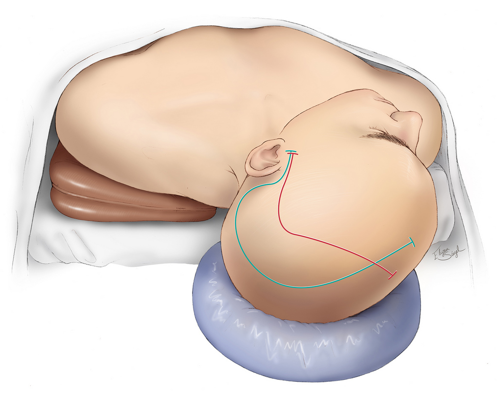
Temporal/Subtemporal Craniotomy
JCM, Free Full-Text

Intraoperative detection of blood vessels with an imaging needle during neurosurgery in humans

Applications of flexible electronics related to cardiocerebral vascular system - ScienceDirect
Craniotomy, Expert Surgeon

Brain surface temperature under a craniotomy
Brain Sciences, Free Full-Text

Craniotomy for acute monitoring of pial vessels in the rodent brain - ScienceDirect

Photothrombotic Middle Cerebral Artery Occlusion in Mice: A Novel Model of Ischemic Stroke

Astrocyte-neuron lactate shuttle plays a pivotal role in sensory-based neuroprotection in a rat model of permanent middle cerebral artery occlusion

PDF] Jugular bulb temperature: comparison with brain surface and core temperatures in neurosurgical patients during mild hypothermia.

Full article: Brain temperature and its role in physiology and pathophysiology: Lessons from 20 years of thermorecording

a) Relationship between temporal change in cerebral blood flow and
Recomendado para você
-
 respuesta del nivel 367 de brain test06 julho 2024
 -
 Results of whole brain analyses in the test scan. a The left parietal06 julho 2024
 -
 Brain Test, Nivel 367, Quiere tener grandes musculos, Explicado Español06 julho 2024
 -
 The genetic architecture of the human cerebral cortex06 julho 2024
 -
 Test your brain Cold Spring Harbor Laboratory06 julho 2024
 -
Day 367 of #stevensnewnormal Turning my focus from this-time-last-year to day. Chuck continues to warrior through and make progress. He.…06 julho 2024
-
 Saatnya mencari cuan! (Brain Test Level 367) - CadeMedia06 julho 2024
 -
 HOME06 julho 2024
 -
:max_bytes(150000):strip_icc()/what-is-serotonin-5189485_color_v1-0cf1021dcefb4410865f4cea18254b5e.jpg) What Is Serotonin?06 julho 2024
What Is Serotonin?06 julho 2024 -
 Gut Microbiome–Brain Alliance: A Landscape View into Mental and06 julho 2024

você pode gostar
-
Márcia Fú com medo do sapo que apareceu na baia 🐸 ! WL pegunta para e06 julho 2024
-
 The Evil Dead American Horror Film Cover Illustration Home Decor06 julho 2024
 -
 trading cards cromo carta pokemon 2012 ingles z - Comprar Cartas Colecionáveis antigas no todocoleccion06 julho 2024
 -
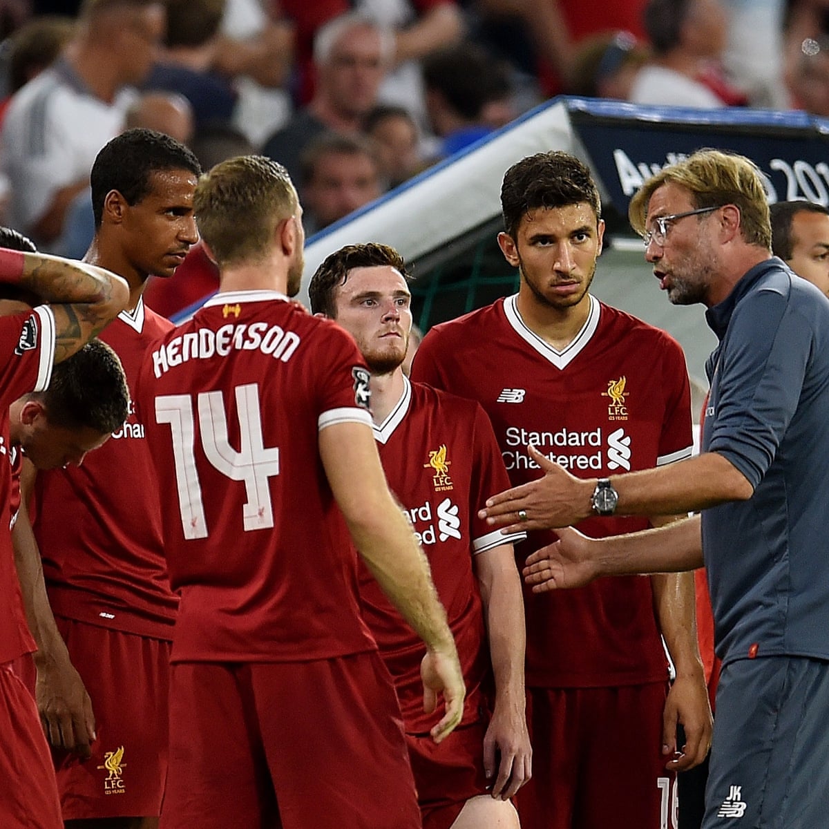 Premier League 2017-18 preview No10: Liverpool, Liverpool06 julho 2024
 -
 Campeonato Paulista 2024: fórmula é aprovada, e grupos estão definidos06 julho 2024
 -
 Pokemon zekrom and reshiram 9806 julho 2024
 -
 V for Vendetta (2005)06 julho 2024
 -
 Ben 10: Ultimate Alien06 julho 2024
 -
/origin-imgresizer.eurosport.com/2020/09/21/2891435-59536708-2560-1440.jpg) Italian Open to host fans from last-16 stage, WTA announces new06 julho 2024
Italian Open to host fans from last-16 stage, WTA announces new06 julho 2024 -
 The New Mutants: what should we expect from the cursed X-Men horror film?, Horror films06 julho 2024
